|
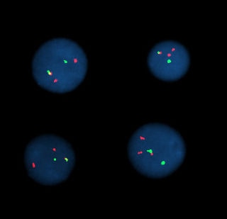
On Tuesday afternoon I learned about tethered cord syndrome, which is a disorder in which tissues limit the movement of the spinal cord. As the child grows the spinal cord will stretch which can lead to nerve damage and pain. Tethered cord often occurs in patients who have spina bifida, a birth defect where the spinal cord does not close properly.  On Thursday I spent the afternoon in the Genetics Lab at Nationwide Children's Hospital. When I arrived I was first taken on a tour by one of the lab genetics counselors. I got to walk through all the different rooms and learn about the different machines and techniques used in each room. While walking through the rooms, I learned was different lab coats need to be worn at each location of the lab to avoid any contamination. Some of the different sections of the lab are tissue, amniotic, and dark. In several of these rooms the pressure and temperature needed to be controlled do that the chromosomes would stick to the glass slides better. Examples of different testing conducted are microarray, whole exome sequencing, gel electrophoresis, FISH, and tests on tissue, blood, and amniotic fluid. Microarrays are used to determine if there are missing or extra pieces of DNA. To do this the patients DNA, and the DNA of an unaffected individual of the same gender is needed. The patients DNA is fluorescently labeled green, and the normal DNA is labeled red. The DNA is then mixed and dropped on a microarray chip. The chip is placed in a laser scanner which will show either yellow, red, or green for each gene. If the gene is yellow, the gene is present. If the gene is red, that means part, or all of it is missing. If it is green, it is a duplication. The last room I toured was the dark room which had to be entered through a revolving darkroom door. Inside the dark room Fluorescence in situ hybridization (FISH) is conducted. The FISH method is used to count how many 13, 18, 21, X, and Y chromosomes there are. To do so probes, DNA strands with a fluorescent label, will glow when the chromosome is present. For example if the patient is suspected of having trisomy 13 the probes that corresponds to the 13th chromosomes will be mixed with the DNA sample. If three green dots appear, the patient has trisomy 13. If there are only 2 green dots then the person has the correct number of chromosome 13s.
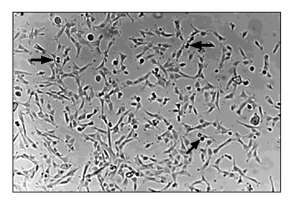
After the tour I observed two lab technicians prepare amniotic fluid samples that will be used for chromosomal analysis. Micropipettes were used to remove cells from the fluid and suspend them in a medium. The mixture was then spread onto four different petri dishes. Two were placed in one fridge, and the other two were placed in a different one in case one would stop working. In about a week the cells would have grown enough to be examined under a microscope to be analyzed. The cells that are not used for testing are then frozen. They can either be flash frozen with liquid nitrogen or simply be frozen. The cells that are flash frozen can be observed, but can no longer grow. Cells that have only been frozen though can theoretically still be grown, even the ones that have been in the freezer since the 1970s.
The last thing I did was look at how karyotypes are created. A karyotype is an organized visual of what a persons chromosomes look like. First several pictures are taken of the chromosomes under the microscope. The pictures need to be clear enough to see each chromosome and each of its bands. Each sister chromatid is then paired up with its match so that all the chromosome can easily viewed at once. 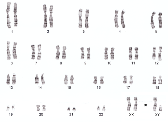
0 Comments
|